

Project
Project
Atherosclerosis and its consequences remain the main cause of mortality in industrialized and developing nations. Despite major advances in treatment, a large number of victims die or are disabled either because the first manifestation is sudden death or an acute cardiovascular event, or because of lack of treatment efficacy, that can be partly caused by the inadequacy of the treatment.
In order to reduce the risk of an acute event, unstable atherosclerosis has to be detected at an early stage of its development. However, the imaging tools that are currently available to detect the spread of atherosclerosis and to predict the associated risk are mainly based on morphological plaque and arterial wall criteria such as calcium scoring or Intima-Media Thickness (IMT). However morphological features only represent the changes in the arterial wall, while it is inflammation that causes plaque evolution. So, obtaining direct, whole body, information on plaque inflammation, including coronary arteries, using target-specific contrast agents would strongly improve the prediction of acute cardiovascular events and thus lead to a better stratification of patients' risk as well as a better fine-tuning of (preventive) treatment.
Furthermore, when an acute event (MI or stroke) has occurred, microcirculation impairment and secondary inflammation processes play an important role in the final lesion size and thus in patient outcome. Having a fast imaging method with contrast agent specific to inflammation and damages to the micro-circulation process would allow us to better adapt the therapy to the individual patients but also to better assess the efficacy of new therapies such as cyclosporine in acute MI.
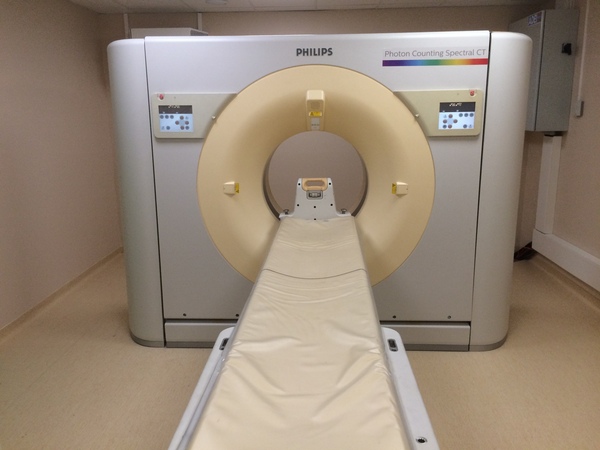
Thus, developing a fully clinically functioning SPCCT modality necessitates developing and merging two technologies:
(a) the acquisition system (mainly through the development of the detector) on one hand
(b) and specific contrast agents on the other hand.
The core objective of this project is therefore to develop and validate a widely accessible, new quantitative and analytical imaging technology combining Spectral Photon Counting Computed Tomography (SPCCT) AND dedicated contrast agents to accurately detect, characterize and monitor neurovascular and cardiovascular disease.
To this end, teams of radiologists and researchers from Lyon are joining forces with other scientists, public institutions and industrialists to carry out this research and enable the development and improvement of this SPCCT as well as the various contrast agents