

KOLOR SPCCT Imaging
KOLOR SPCCT Imaging
ERC Starting Grant project
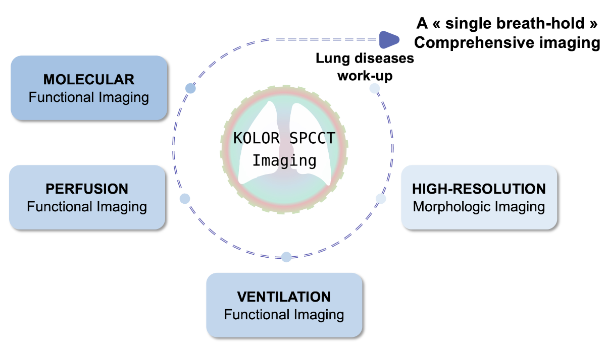
X-ray computed tomography (CT) stands as the cornerstone of pulmonary imaging due to its higher spatial resolution, convenience, availability, and faster acquisition time compared to other imaging methods such as magnetic resonance imaging and nuclear imaging. However, it only provides morphological characterization, which is not fully suited to pulmonary diseases that involve a complex combination of respiratory, vascular, and inflammatory dysfunctions. Their diagnosis requires both morphological and functional analysis of ventilation, perfusion, and molecular biomarkers. Thus, the standard of care relies on a multimodal diagnostic assessment involving scintigraphy, positron emission tomography, and tissue biopsy. This presents three main drawbacks: either the assessment is not precise enough, it is invasive, or it is time-consuming while worsening the patient's prognosis.
Spectral Photon-Counting Computed Tomography (SPCCT) not only capitalizes on all the advantages of morphological CT imaging but also offers an advanced imaging method known as K-Edge imaging. This method allows the specific and quantitative identification of one or more atoms simultaneously in tissue, enabling simultaneous functional imaging of independent or interactive processes. However, K-Edge color imaging is still limited by its low sensitivity and the scarcity of tracers for potential human use, and thus has not been put into practice yet.
By combining medical imaging, respirology, chemistry, and physics, KOLOR SPCCT imaging will bridge the gap between morphological and functional imaging in a single breath. To achieve this goal, the project proposes to:
- Develop K-Edge color imaging: create a highly sensitive dedicated imaging tool.
- Diagnose pulmonary diseases: provide "single-breath" imaging of ventilation and perfusion in animal models.
- Predict pulmonary diseases: ensure "single-breath" monitoring of the inflammatory molecular load in animal models.
KOLOR SPCCT imaging will provide unprecedented insights into ventilation, perfusion, molecular inflammatory response, and their interactions. It will develop K-Edge color imaging to provide specific and high-resolution quantitative imaging using both non-specific and specific tracers on key pulmonary applications in animal models: pulmonary embolism, cancer, and fibrosis. This will lead to a paradigm shift in the diagnosis and prognosis of pulmonary diseases, enabling earlier and more accurate diagnosis for a greater chance of patient survival.