Contrast Agents & K-Edge Imaging
Contrast Agents & K-Edge Imaging
Moving forward contrast agents’ production and PoC: this transversal project addresses one of the main challenges for molecular imaging agents, i.e. the scale-up process from lab chemistry to production to enabling successively proof-of-concept (PoC) studies, pre-clinical and clinical trials. Whereas today, the imaging agent market is dominated by contrast agents (65% of the market for 35% to SPECT/TEP tracers) for cardiovascular and brain
imaging, development of new targeted imaging agents is anticipated. It is expected to continue its increase ($10.4 million of global pharmaceutics in 2010, and estimated $14.1 million in 2015). The main challenge is however to keep the application spectrum large enough for sales turnover. Imaging agents are also envisaged as new biomarkers and test companions for drug clinical trials, enabling faster transfer of innovative treatment to patients by a more personalized approach. Production and PoC will therefore be an essential step, but only for a limited number of candidates. The first step, human cell culture test will be obtained with a minimal production concern, but testing in small animals implies a scale-up for synthesis, with various issues depending on the platform (macromolecule,nanoparticles) and on the target moiety. This task will require complementary competencies for selection of relevant animal models for human receptor expressions (molecular expression is a dynamic process during disease evolution, and its relevance to diagnosis of human diseases is often not direct), 2/ for biological assays, 3/ for initial biodistribution and pharmaco-kinetics to determine dose and time-window for optimal concentration at target, and 4/ for optimized imaging and interventional protocols as well as minimal toxicity. After the termination of the project, effective translational research with first in-man evaluations can be anticipated in the neuro and cardiovascular diseases.
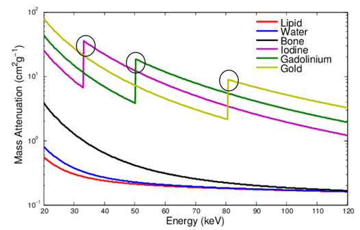
SPCCT allows a new mapping called "K-Edge". The K-Edge is the binding energy of an electron on the electronic layer K with its nucleus. The photons having an energy equal to this energy will be attenuated, creating a distinguishable attenuation peak. The energy peaks related to each energy layer are also present but their energy being lower, they do not fit in the detection spectrum of photon counting detectors because these photons will be strongly attenuated by the human body.
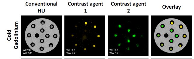
From this point of view, the SPCCT, thanks to K-Edge imaging, makes it possible to carry out even more research into contrast agents, such as the differentiation of two contrast agents with high K-Edges as Gold & Gadolinium that was previously impossible in conventional CT:
K-Edge Imaging
PCDs also allow a new mapping called "K-Edge". The K-Edge is the binding energy of an electron on the K electronic layer with its nucleus. Photons with an energy equal to this energy will be attenuated, creating a distinguishable attenuation peak. The energy peaks linked to each energy layer are also present but their energy being much lower, they do not fit in the detection spectrum of PCDs because these photons will be strongly attenuated by the human body.
This type of imaging requires the measurement of at least three energy bins which is not feasible with actual clinical scanners. Contrast agents with high K-Edge energies as Gadolinium or Gold can be discriminated with this “K-Edge Imaging":
Conventional imaging does not allow discrimination between these two contrast agents unlike K-Edge imaging. This new mapping opens up a new diagnostic possibility of diagnosis. Indeed, by imagining that we could "tag" a disease thanks to a contrast product, this imaging mode would allow a more advanced diagnosis. This imaging method would allow a more advanced and safer diagnosis.