Photon Counting Detectors
Photon Counting Detectors
The scanner is the most widely used imaging method in the world, transforming patient care. Scanners provide a black-and-white image of the human body but bring its disadvantages with higher X-ray dose than conventional X-ray imaging and poor differentiation of soft tissue. It too frequently requires complementary medical exams such as MRI in order to confirm diagnosis.
The development of the SPCCT is taking part of this medical context. Based on new detectors developed by Philips, it allows a direct and unitary counting of photons and their classification by energy level resulting in a higher spatial resolution, a decrease in the dose received by the patient and a spectral analysis of body elements.
This technological innovation should lead to a better lesion characterization avoiding sampling and continue to expand Computed Tomography to screening, due to the reduction of radiation doses
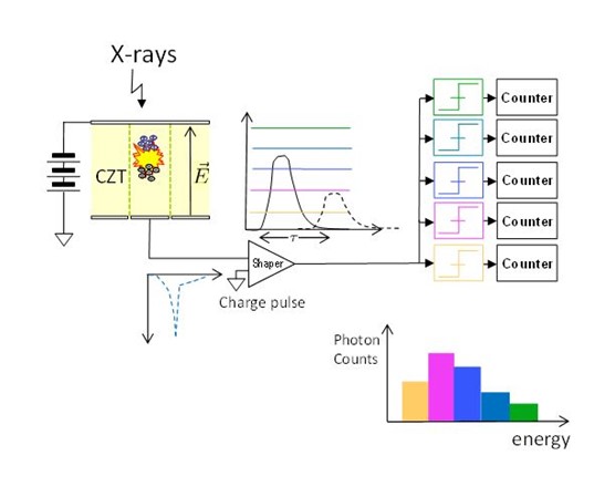
Photon Counting Detectors
Unlike the integrated detectors used in conventional scanners, PCDs are made of CZT (Cadium Telluride Zinc) semiconductors. The first layer of semiconductor of thickness from 2 to 3mm, allows, by absorbing a photon, to form electron-hole pairs. These detectors are connected to an electronic system (ASIC) composed of parallel channels allowing successively to model the signal and to amplify it so that it is sorted by energy level. The photons are then discriminated in 2 to 8 energy levels chosen beforehand. Operation of the detectors is summarized in the schematic below: