

Benefits
Benefits
Spatial Resolution
Facilitating vascular and perfusion imaging by enabling higher spatial resolution.
The spatial resolution offered by SPCCT is limited by the range of the photo-electron produced by an absorbed x-ray photon and in a practical system by the size of the pixelated anodes on the sensor crystal and the focal spot of the X-Ray Tube. This former range is under 100 μm for the diagnostic energy range, producing signal charge clouds of about that size. Consequently, if reasonable energy resolution is desired the pixel size should be chosen significantly bigger than this so as to seldom split signal charge clouds.
Even with this requirement the spatial resolution of our Animal scanner improved the conventional CT resolution two fold, without the need for comb spatial filters or other devices that achieve high resolution at the cost of increased dose.
The Animal scanner demonstrated 24 line-pairs/cm at regular scanning dose rates. This high resolution offers new opportunities to examine the vascular system in vivo and to diagnose a variety of conditions that were not previously accessible.
The resolution capability will be combined with the material decomposition sensitivity to determine first pass and perfusion in the vascular system, for example with Gd or I blood pool markers, or with complexed pharmaceuticals utilizing these markers.
This resolution capability could be extended for Oncology: the high resolution and the material decomposition sensitivity are also useful for detecting pathology of specific organ systems, such as liver, kidneys etc; and for detecting physiopathological process such as angiogenesis and for specifically active oncology marker.
Electronic Noise
As explained in the part “Photon Counting Detectors” , photons are sorted in 3 to 8 energy levels. Be able to set these thresholds allows to get rid of the electronic noise by taking into account only the photons above the first energy level.
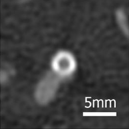
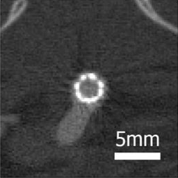
Sigovan, M. et al. “Initial Experience in Improving Stent Analysis and Intra Stent Lumen Assessment Using Spectral Photon Counting CT and K-Edge Imaging”. In RSNA; Chicago, United States, 2016
Thanks to Photon Counting Detectors’ properties, SPCCT images have a better in contrast resolution. The lowest energy photons, which bring the most contrast information (strong photoelectric effect), have the same weight as those of high energy contrary to indirect detection detectors
Dose
All the advantages of these PCDs lead either to a better image quality for the same dose received by the patients allowing to improve the diagnoses or to an equivalent if not superior image quality for a reduced dose received by the patient with the SPCCT.