Innovative Cardiovascular Imaging Technology
Technology
Context
Ct-Scan is the most widely used imaging method in the world, transforming patient care. Scanners provide a black-and-white image of the human body, with the limitation that too many samples are taken to confirm a diagnosis. The objective is to develop a new generation of scanners combined with the development of new contrast agents. The expected gains for patients and the healthcare system are considerable, particularly in oncology and cardiology, which account for over 20% of healthcare costs.
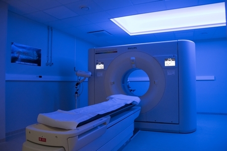
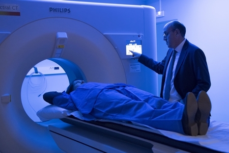
SPCCT : What is it ?
The innovation of the SPCCT is based on a different detector technology allowing direct and unitary counting of photons and their classification by energy level. This leads to significant increase in spatial resolution of the system, a decrease for X-ray dose receive by patients and a spectral analysis of the elements crossed by the X-rays and will thus make it possible to pass from black and white scanning to color scanning. It should therefore make it possible to avoid a certain number of biopsies by better characterizing the lesions in a non-invasive manner. Moreover, by a substantial reduction of the X-ray dose, it could allow to extend to medical imaging and to CT the screening campaigns currently limited to mammography, in order to evaluate the potential risk of stroke or myocardial infarction, or lung cancer with a precision of the order of a tenth of a mm.
Which applications?
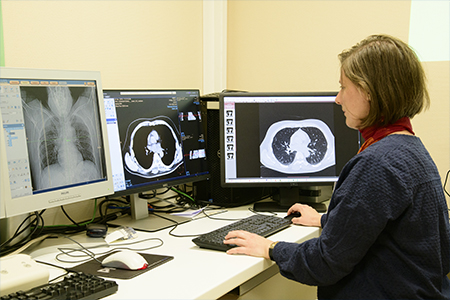
The field of application of this new technique concerns all medical fields. For example, it should eventually allow the detection and measurement of the activity of atherosclerotic plaques or tumors without the use of radioelements, i.e., without the need for the continuous production of radioactive molecules by radio pharmacies. This will allow patients to benefit more widely from the possibilities offered by molecular imaging in screening, lesion identification and analysis of their response to treatment, especially since, after validation, this method should be easily transposable to clinical scanner imaging insofar as the modification concerns "only" the detector and not all the components of the current machines.
It should therefore make it possible to avoid a certain number of biopsies by better characterizing the lesions in a non-invasive manner. Moreover, by a substantial reduction in the dose of X-rays, it could allow the extension of screening campaigns currently limited to mammography to medical imaging and CT scans, in order to evaluate the potential risk of stroke or myocardial infarction, or lung cancer with an accuracy of a tenth of a mm.
The long-term strategy is to establish Spectral CT as a new clinical imaging modality in humans.